|
Overview
 A person with flat feet (fallen arches) has low arches or no arches at all. Most cases don't cause problems and treatment isn't usually needed. The arch, or instep, is the inside part of the foot that's usually raised off the ground when you stand, while the rest of the foot remains flat on the ground. Most people have a noticeable space on the inner part of their foot (the arch). The height of the arch varies from person to person. Causes Abnormal development of the foot, producing Pes Planus, may be due to neurological problems, eg cerebral palsy, polio. Bony or ligamentous abnormalities, eg tarsal coalition (fusion of tarsal bones), accessory navicular bone. A small proportion of flexible flat feet do not correct with growth. Some of these may become rigid if the Pes Planus leads to bony changes. Symptoms Having flat feet can be painless and is actually normal in some people. But others with flat feet experience pain in the heel or arch area, difficulty standing on tiptoe, or have swelling along the inside of the ankle. They may also experience pain after standing for long periods of time or playing sports. Some back problems can also be attributed to flat feet. Diagnosis If you notice that your feet are flat, but you?re not really experiencing any pain, then you?re probably okay to go without a visit to the podiatrist (unless, of course, you have a lack of feeling in your foot). You can schedule a hair appointment instead, or maybe see a movie. However, once painful symptoms start to appear, it?s better to skip the hirsute (or cinematic) experience and go see your foot doctor. Your podiatrist will likely make the diagnosis by examining your foot visually, asking about symptoms you may be experiencing, and may test your muscle strength. You may be asked to stand on your toes (in a ballerina pose, if you prefer, although that?s certainly not required), or walk around the examining room, and you may need to show the podiatrist your shoes. He or she may comment on your excellent taste in footwear, but is more likely to check your shoes for signs of wear that may indicate fallen arches. Your podiatrist may recommend X-rays, a CT scan or an MRI in order to get a look at the interior of your foot, although the best diagnosis usually comes from the doctor?s own in-person examination. arch support inserts reviews Non Surgical Treatment The typical treatment for pain from fallen arches is an arch insert. While many people experience dramatic pain relief from this, others continue to suffer from chronic achy feet despite the arch support. The problem with this approach is that it does not do anything to strengthen the weak ligaments that may be at the root of the problem and, thus, does not alleviate the chronic pain that people with this condition experience. Another standard practice of modern medicine is to use steroids or to prescribe anti-inflammatory medications. However, in the long run, these treatments do more damage than good. Cortisone shots and anti-inflammatory drugs have been shown to produce short-term pain benefit, but both result in long-term loss of function and even more chronic pain by actually inhibiting the healing process of soft tissues and accelerating cartilage degeneration. Plus, long-term use of these drugs can lead to other sources of chronic pain, allergies and leaky gut syndrome. Surgical Treatment  A combination of surgical procedures can be used to reconstruct the flatfoot. Generally, these procedures can be separated into those that correct deformities of the bones and those that repair ligaments and tendons. Your orthopaedic surgeon will choose the proper combination of procedures for your foot. Surgery of the foot can be performed under regional anesthesia, which is numbing the foot and ankle with a nerve or spinal block, or general anesthesia, which may require a breathing tube. A nerve block is often placed behind the knee to reduce pain after surgery. Prevention It?s time to take a long hard look at what?s in your closet. Now is the time to toss out shoes that are well worn. You also need to say good-bye to thin-soled shoes that offer zero arch support. If you?re overweight, fallen arches may be a sign the universe is trying to tell you something. You need to lose weight, and odds are, fallen arches are but one of many physical discomforts you are experiencing. Overview
Leg shortening is employed when LLD is severe and when a patient has already reached skeletal maturity. The actual surgery is called an osteotomy , which entails the removal of a small section of bone in the tibia (shinbone) and sometimes the fibula as well, resulting in the loss of around an inch in total height. Leg lengthening is a difficult third option that has traditionally had a high complication rate. Recently, results have improved somewhat with the emergence of a technique known as callotasis , in which only the outer portion of the bone (the cortex ) is cut, (i.e. a corticotomy ). This allows the bone to be more easily lengthened by an external fixation device that is attached to either side of the cut bone with pins through the skin. The ?ex-fix,' as it is sometimes called, is gradually adjusted by an orthopaedic surgeon, and healing can occur at the same time that the leg is being distracted , or lengthened over time. Unlike epiphysiodesis, leg lengthening procedures can be performed at almost any skeletal or chronological age.  Causes An anatomical short leg is due to several orthopedic or medical condition mechanisms. Often, one leg simply stops growing before the other one does and is called ?congenital?. We often see mother-daughters or father-sons who exhibit virtually the same degree of shortness on the same side. Often it is not known why this occurs, but it seems to account for approximately 25% of the population demonstrating a true LLD. Other causes of a true LLD include trauma or broken bones, surgical repair, joint replacement, radiation exposure, tumors or Legg-Calves-Perthes disease. Symptoms The patient/athlete may present with an altered gait (such as limping) and/or scoliosis and/or low back pain. Lower extremity disorders are possibly associated with LLD, some of these are increased hip pain and degeneration (especially involving the long leg). Increased risk of: knee injury, ITB syndrome, pronation and plantar fascitis, asymmetrical strength in lower extremity. Increased disc or vertebral degeneration. Symptoms vary between patients, some patients may complain of just headaches. Diagnosis Asymmetry is a clue that a LLD is present. The center of gravity will shift to the short limb side and patients will try to compensate, displaying indications such as pelvic tilt, lumbar scoliosis, knee flexion, or unilateral foot pronation. Asking simple questions such as, "Do you favor one leg over the other?" or, "Do you find it uncomfortable to stand?" may also provide some valuable information. Performing a gait analysis will yield some clues as to how the patient compensates during ambulation. Using plantar pressure plates can indicate load pressure differences between the feet. It is helpful if the gait analysis can be video-recorded and played back in slow motion to catch the subtle aspects of movement. Non Surgical Treatment In some circumstances, the physician will recommend a non-surgical form of treatment. Non-surgical treatments include orthotics and prosthetics. Orthotics are a special type of lift placed in or on a shoe that can be used in the treatment of leg length discrepancies between two and six centimeters. In pediatric patients who have large discrepancies and are not good candidates for other treatment forms, prosthetics can be helpful.  what are shoe lifts? Surgical Treatment Leg shortening is employed when LLD is severe and when a patient has already reached skeletal maturity. The actual surgery is called an osteotomy , which entails the removal of a small section of bone in the tibia (shinbone) and sometimes the fibula as well, resulting in the loss of around an inch in total height. Leg lengthening is a difficult third option that has traditionally had a high complication rate. Recently, results have improved somewhat with the emergence of a technique known as callotasis , in which only the outer portion of the bone (the cortex ) is cut, (i.e. a corticotomy ). This allows the bone to be more easily lengthened by an external fixation device that is attached to either side of the cut bone with pins through the skin. The ?ex-fix,' as it is sometimes called, is gradually adjusted by an orthopaedic surgeon, and healing can occur at the same time that the leg is being distracted , or lengthened over time. Unlike epiphysiodesis, leg lengthening procedures can be performed at almost any skeletal or chronological age. Overview
 The most common cause of heel pain is plantar fasciitis which is commonly referred to as a heel spur. Plantar fascia is a broad band of fibrous tissue which runs along the bottom surface of the foot, from the heel to the toes. Plantar fasciitis is a condition in which the plantar fascia is inflamed. This condition can be very painful and cause a considerable amount of suffering. Causes Heel pain has many causes. Heel pain is generally the result of faulty biomechanics (walking gait abnormalities) that place too much stress on the heel bone and the soft tissues that attach to it. The stress may also result from injury, or a bruise incurred while walking, running, or jumping on hard surfaces; wearing poorly constructed footwear (such as flimsy flip-flops); or being overweight. Symptoms The symptoms of plantar fasciitis include pain along the inside edge of the heel near the arch of the foot. The pain is worse when weight is placed on the foot especially after a long period of rest or inactivity. This is usually most pronounced in the morning when the foot is first placed on the floor. This symptom called first-step pain is typical of plantar fasciitis. Prolonged standing can also increase the painful symptoms. It may feel better after activity but most patients report increased pain by the end of the day. Pressing on this part of the heel causes tenderness. Pulling the toes back toward the face can be very painful. Diagnosis To arrive at a diagnosis, the foot and ankle surgeon will obtain your medical history and examine your foot. Throughout this process the surgeon rules out all the possible causes for your heel pain other than plantar fasciitis. In addition, diagnostic imaging studies such as x-rays or other imaging modalities may be used to distinguish the different types of heel pain. Sometimes heel spurs are found in patients with plantar fasciitis, but these are rarely a source of pain. When they are present, the condition may be diagnosed as plantar fasciitis/heel spur syndrome. Non Surgical Treatment Initially, treatment will consist of adding support to the foot, including better shoes and an over-the-counter arch supports and/or insoles; resting from the sport or activity that aggravates the problem; stretching the calf and arch muscles; taking anti-inflammatory; and using ice and massage to reduce inflammation. You can ice and message your muscles simultaneously by freezing a water bottle filled with water and using it to massage your foot by rolling it underneath your foot for five to 10 minutes at least two times per day. It is not unusual for symptoms of plantar fasciitis to persist for six to 12 months despite treatment. Surgical Treatment It is rare to need an operation for heel pain. It would only be offered if all simpler treatments have failed and, in particular, you are a reasonable weight for your height and the stresses on your heel cannot be improved by modifying your activities or footwear. The aim of an operation is to release part of the plantar fascia from the heel bone and reduce the tension in it. Many surgeons would also explore and free the small nerves on the inner side of your heel as these are sometimes trapped by bands of tight tissue. This sort of surgery can be done through a cut about 3cm long on the inner side of your heel. Recently there has been a lot of interest in doing the operation by keyhole surgery, but this has not yet been proven to be effective and safe. Most people who have an operation are better afterwards, but it can take months to get the benefit of the operation and the wound can take a while to heal fully. Tingling or numbness on the side of the heel may occur after operation. bestshoelifts Prevention  It is not always possible to prevent heel pain, but there are measures you can take to help avoid further episodes. Being overweight can place excess pressure and strain on your feet, particularly on your heels. This increases the risk of damaging your feet and heels. If you are overweight, losing weight and maintaining a healthy weight by combining regular exercise with a healthy, balanced diet can be beneficial for your feet. You can calculate your body mass index (BMI) to find out whether you are a healthy weight for your height and build. To work out your BMI, divide your weight in kilograms by your height in metres squared. A BMI of less than 18.5 means that you are underweight, 18.5-24.9 means that your weight is healthy, 25-29 means that you are overweight, 30-40 means that you are obese, over 40 means that you are morbidly obese. You can also use the BMI healthy weight calculator to work out your BMI. Overview
 Morton neuroma (interdigital neuroma), first described in 1876, is a perineural fibrosis and nerve degeneration of the common digital nerve. Morton neuroma, or Morton's neuroma, is not a true neuroma, although it results in neuropathic pain in the distribution of the interdigital nerve secondary to repetitive irritation of the nerve. The most frequent location is between the third and fourth metatarsals (third webspace). Other, less common locations are between the second and third metatarsals (second webspace) and, rarely, between the first and second (first webspace) or fourth and fifth (fourth webspace) metatarsals. Morton neuroma (interdigital neuroma), first described in 1876, is a perineural fibrosis and nerve degeneration of the common digital nerve. Morton neuroma, or Morton's neuroma, is not a true neuroma, although it results in neuropathic pain in the distribution of the interdigital nerve secondary to repetitive irritation of the nerve. The most frequent location is between the third and fourth metatarsals (third webspace). Other, less common locations are between the second and third metatarsals (second webspace) and, rarely, between the first and second (first webspace) or fourth and fifth (fourth webspace) metatarsals.Causes Inappropriate footwear is one of the principle causes of Morton?s neuroma. Toe spring and tapering toe boxes are the most problematic shoe design features that contribute to this health problem. Morton?s neuroma occurs when one of your nerves is stretched and pinched, which happens with great frequency in people who wear shoes incorporating these design features. A professional shoe fitting should always be sought if you are struggling with neuroma-related symptoms. Symptoms You may initially experience a tingling sensation in the space between your toes, which gets worse over time. This leads to cramp in your toes and a sharp shooting or burning pain on the ball of your foot or at the base of your toes. The pain is often worse when walking or wearing shoes that press on the affected area. This is caused by irritation of the nerve between your toe bones (metatarsal bones). The tingling will eventually lead to pain, which can get worse over time. You may also experience cramping of your toes. The pain is usually felt as a sharp shooting or burning pain on the ball of the foot or at the base of the toes, which is often made worse when you're walking. Some people with Morton's neuroma feel anxious about walking or even placing their foot on the ground. The pain is likely to be more intense if you wear tight shoes, so wearing shoes that have more room in the toe area can help. Rubbing your foot may also lessen the pain. Diagnosis X-rays of your affected foot will not show a neuroma, as neuromas are made up of soft tissue. X-rays may be helpful, however, in helping rule in osteoarthritis or a stress fracture as the cause of your symptoms. Ultrasonography and MRI are sometimes used to help diagnose neuromas, although they are often ineffective. The clinical diagnosis by a foot care expert with experience treating this health problem is usually the most effective way to diagnose neuromas. Your podiatrist will attempt to duplicate your neuroma symptoms by pressing on the involved nerve at various points, and he or she may try to cause a clicking of your nerve that indicates nerve enlargement. Non Surgical Treatment Once a diagnosis is obtained, it is essential to begin treatment immediately. Your podiatric physician will advise you on the most effective means. If caught early enough, good foot care, shoes that fit properly, and/or orthoses may eliminate the need for any further intervention. Other conservative measures might include oral non-steroidal anti-inflammatory medication (NSAIDS), physical therapy, ultrasound or other non-invasive measures. If that doesn?t work, your podiatric physician might use injectable steroids, and/or a local anesthetic around the neuroma to reduce inflammation and pain. Many patients report relief after these measures are taken.  Surgical Treatment Surgery to excise the neuroma is usually performed under general anaesthetic in a day surgery facility. After surgery you will have to keep your foot dry for two weeks. Generally neuroma surgery allows for early weight bearing and protection in some type of post op shoe gear. Some neuromas may reoccur, but this is rare. Most studies on patient satisfaction after neuroma surgery show approximately 90% reduction of pain and about 85% of all patients rated the overall satisfaction with the results as excellent or good. There are two unique variations of leg length discrepancies, congenital and acquired. Congenital indicates that you are born with it. One leg is anatomically shorter compared to the other. As a result of developmental phases of aging, the human brain senses the stride pattern and identifies some variation. Our bodies typically adapts by tilting one shoulder to the "short" side. A difference of less than a quarter inch isn't grossly uncommon, require Shoe Lifts to compensate and generally won't have a serious effect over a lifetime.
 Leg length inequality goes largely undiagnosed on a daily basis, yet this issue is simply fixed, and can eradicate numerous incidents of lower back pain. Treatment for leg length inequality usually involves Shoe Lifts. These are affordable, commonly being less than twenty dollars, in comparison to a custom orthotic of $200 or higher. Differences over a quarter inch can take their toll on the spine and should probably be compensated for with a heel lift. In some cases, the shortage can be so extreme that it requires a full lift to both the heel and sole of the shoe. Back ache is the most common ailment afflicting people today. Around 80 million people are afflicted by back pain at some stage in their life. It is a problem which costs businesses huge amounts of money each year due to time lost and output. New and more effective treatment methods are continually sought after in the hope of minimizing the economic impact this issue causes.  Men and women from all corners of the earth experience foot ache due to leg length discrepancy. In these types of cases Shoe Lifts can be of immense help. The lifts are capable of relieving any discomfort in the feet. Shoe Lifts are recommended by numerous qualified orthopaedic orthopedists. So that you can support the body in a well balanced fashion, your feet have a crucial part to play. Inspite of that, it can be the most overlooked zone of the human body. Many people have flat-feet which means there is unequal force placed on the feet. This will cause other body parts including knees, ankles and backs to be affected too. Shoe Lifts ensure that ideal posture and balance are restored.  Overview OverviewHammer toe is most common in women, and a big part of this is poor shoe choices, which are a big factor in the development of many foot problems. Tight toe boxes and high heels are the biggest culprits. Genetics certainly plays a role in some cases of hammertoes, as does trauma, infection, arthritis, and certain neurological and muscle disorders. Most cases of contracted toes are associated with various biomechanical abnormalities in how a patient walks. This causes the muscles and tendons to be used excessively or improperly, which deforms the toes over time. Causes Hammer toe is often caused by wearing shoes that do not fit properly. If shoes are too small either in length or width, then the toes are held in a shortened position for long periods and the muscles eventually shorten and pull the toes into the bent position. Alternatively it can be caused by overactivity in the extensor digitorum dongus muscle (right) and a weakness in the counteracting muscle under the foot, such as flexor digitorum longus. Sometimes it can be a congenital condition, meaning it is present from birth. It is also more common in those with arthritis in the foot or diabetes.  Symptoms SymptomsWell-developed hammertoes are distinctive due to the abnormal bent shape of the toe. However, there are many other common symptoms. Some symptoms may be present before the toe becomes overly bent or fixed in the contracted position. Often, before the toe becomes permanently contracted, there will be pain or irritation over the top of the toe, particularly over the joint. The symptoms are pronounced while wearing shoes due to the top of the toe rubbing against the upper portion of the shoe. Often, there is a significant amount of friction between the toe and the shoe or between the toe and the toes on either side of it. The corns hammertoes may be soft or hard, depending on their location and age. The affected toe may also appear red with irritated skin. In more severe cases, blisters or open sores may form. Those with diabetes should take extra care if they develop any of these symptoms, as they could lead to further complications. Diagnosis The treatment options vary with the type and severity of each hammer toe, although identifying the deformity early in its development is important to avoid surgery. Your podiatric physician will examine and X-ray the affected area and recommend a treatment plan specific to your condition. Non Surgical Treatment Wear sensible shoes. If you don?t want to have surgery to fix your hammertoe, use non-medicated padding along with proper shoes made with a wider and deeper toe box to accommodate your foot?s shape. Ensuring your shoes have a good arch support can slow the progression of the condition as well. Use a pumice stone. The corn or callus that forms on top of the hammertoe can cause discomfort when you wear shoes. Treat the corn by using a file or pumice stone to reduce its size after a warm bath, then apply emollients to keep the area softened and pliable. Use silicone or moleskin padding on top of the area when wearing shoes. Do foot exercises. Theoretically, exercises like extending, then curling the toes, splaying the toes, and moving the toes individually may help prevent the digital contracture that causes hammertoe. Try these suggestions and see what works best for you. Surgical Treatment If conservative measures fail to provide relief, or if your hammertoe is in advanced stages with rigidity and a significant amount of pain, surgery may be required. Some patients also require surgery if they have open sores or wounds related to their hammertoe. For patients who also suffer from bunions, a combined procedure may be appropriate, addressing both conditions within the same surgery. Recovery time will vary from patient to patient, depending on the extent of the surgical repair and other conditions that may also be present. 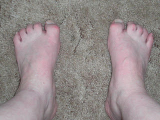 Overview OverviewHammer, mallet, and claw toes have distinctive differences that can assist you in determining what kind of toe problem you are dealing with. All three conditions deal with toes that are curved into abnormal positions, which possibly look strange and may cause pain. Typically, the big toe is not affected by these problems. A Hammer toes tends to bend downward at the center of a toe joint. It generally affects your second toe. The affliction causes the center of your toe to rise and is often accompanied with a bony lump. Causes The main cause of hammer toe is poorly fitted and/or poorly designed footwear. Any footwear that is too tight in the toe box, especially high-heeled shoes, can push the toes forward, crowding one or more of them into a space that is not large enough to allow the toes to lie flat and spread as they should. Other causes include the following. Changes in foot anatomy. Sometimes the metatarsal bones in the ball of the foot can ?drop,? creating a situation in which the toes do not make contact with the surface of the shoe. The toes may then contract at one or both of the joints to re-establish contact with the surface. Traumatic injuries in which toes are jammed or broken. Diabetic neuropathy. This can cause abnormal foot biomechanics due to nerve and/or muscle damage. Damage to nerves and muscles from other conditions, such as arthritis or stroke. Heredity.  Symptoms A hammertoe causes you discomfort when you walk. It can also cause you pain when trying to stretch or move the affected toe or those around it. Hammertoe symptoms may be mild or severe. Mild Symptoms, a toe that is bent downward, corns or calluses. Severe Symptoms, difficulty walking, the inability to flex your foot or wiggle your toes, claw-like toes. See your doctor or podiatrist right away if you develop any of these symptoms. Diagnosis Most health care professionals can diagnose hammertoe simply by examining your toes and feet. X-rays of the feet are not needed to diagnose hammertoe, but they may be useful to look for signs of some types of arthritis (such as rheumatoid arthritis) or other disorders that can cause hammertoe. If the deformed toe is very painful, your doctor may recommend that you have a fluid sample withdrawn from the joint with a needle so the fluid can be checked for signs of infection or gout (arthritis from crystal deposits). Non Surgical Treatment A person with hammer toes will be asked to practice some exercises for their toes to regain average structure and movement. The exercises usually involve stretching and strengthening their toes. The person may attempt to pick things up off the floor using only their toes. They may also stretch their toes on a regular basis by hand to ease them into straightening out. Another example of a physical exercise specifically for a person's toes involves crumpling a towel with the toes. The towel can lie underneath the person's feet and the person can use their toes to scrunch up the towel as they perform simple tasks such as reading a book or watching television. Surgical Treatment he basis for hammer toe surgery most often involves removing s portion of bone within the toe, to reduce the joint contracture. Depending on the direction the toe is deviated, soft tissue procedures may be necessary along with pinning the toe with a surgical wire.  Prevention There should be at least one-half inch between the tip of your longest toe and the front of the shoe. Never buy shoes that feel tight and expect them to stretch with wearing. If you have prominent areas on your feet such as hammertoes and bunions, avoid shoes with a lot of stitching or multiple pieces of fabric, as these stitched areas tend not to stretch to accommodate various toe deformities.
Overview
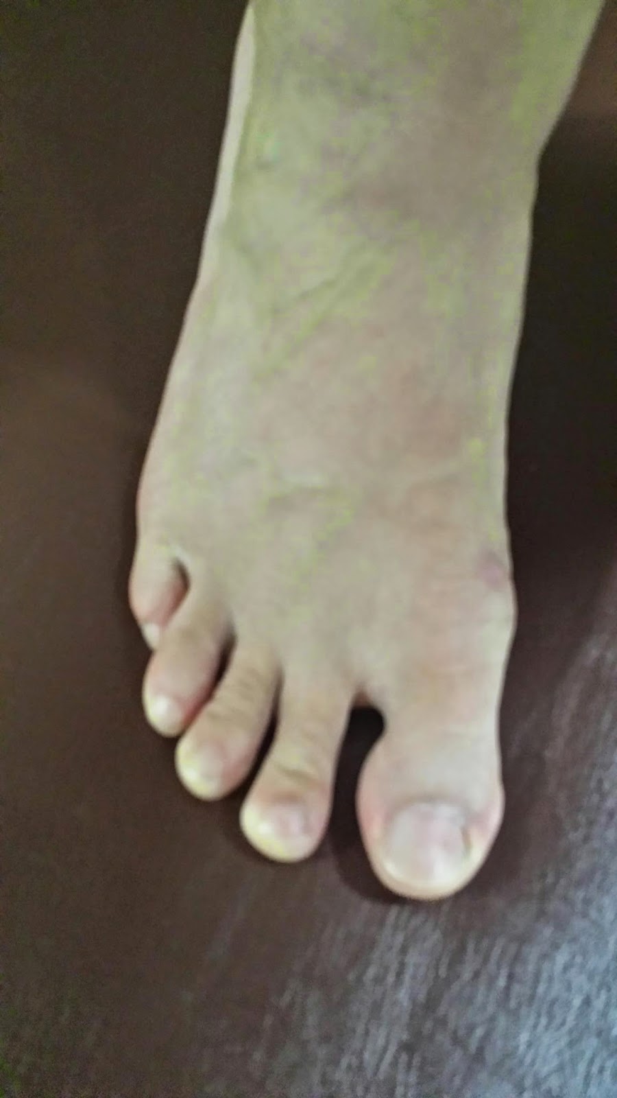
 A bunion is an enlargement of bone or tissue around the joint at the base of the big toe. The big toe may turn toward the second toe. The tissues around the joint may be swollen and tender. A bony bump at the base of the little toe is called a bunionette or tailor's bunion. The little toe also bends inward, and the joint swells or enlarges. A bunion is an enlargement of bone or tissue around the joint at the base of the big toe. The big toe may turn toward the second toe. The tissues around the joint may be swollen and tender. A bony bump at the base of the little toe is called a bunionette or tailor's bunion. The little toe also bends inward, and the joint swells or enlarges.Causes Bunions can be caused by improper footwear. Genetics. Foot injuries. Congenital deformities. Medical conditions such as arthritis. Stress on feet. Bunions are mainly caused by genetics. The bunion itself is not inherited, but the person?s hereditary foot type and gait pattern makes them more prone to developing bunions.You can also begin to develop bunionsby wearing shoes that are too tight or too small. When you wear shoes of this nature, your toes are squeezed together. Bunions are not caused by crowding of the toes, but wearing tight shoes can worsen the condition and cause symptoms to appear sooner. Some people are born with birth defects that put them at higher risk for developing bunions. Symptoms Signs and symptoms of a bunion include the base of the big toe is swollen and sticks out. The big toe is often bent towards the other toes, and sometimes the second toe is pushed to overlap the third toe. Skin around the big toe joint is red and sore. Thickened skin at the base of the big toe. Pain in the big toe or foot. Wearing shoes is painful. Pain or difficulty when walking. Diagnosis Generally, observation is adequate to diagnose a bunion, as the bump is obvious on the side of the foot or base of the big toe. However, your physician may order X-rays that will show the extent of the deformity of the foot. Non Surgical Treatment Patients should immediately cease using improperly fitted shoes. Footwear selection should have a wide and roomy toebox to accommodate the full width of the foot. If the problem is the over-pronation, the patient should be fitted with orthotics and can expect a slow recovery from pain over a period of months. Orthotics will not cause the physical deformity to regress, but will simply arrest any further progression and likely stop the pain. It is important to note however, that when bunions are severe and require surgery, the bunion can be corrected, but will develop again unless the root cause of over-pronation is corrected. If over-pronation is the root cause, orthotics will still be necessary.  Surgical Treatment When a surgeon cuts and repositions a bone, it is referred to as an osteotomy. There are two basic techniques used to perform an osteotomy to realign the first metatarsal. In some cases, the far end of the bone is cut and moved laterally (called a distal osteotomy). This effectively reduces the angle between the first and second metatarsal bones. This type of procedure usually requires one or two small incisions in the foot. Once the surgeon is satisfied with the position of the bones, the osteotomy is held in the desired position with one, or several, metal pins. Once the bone heals, the pin is removed. The metal pins are usually removed between three and six weeks following surgery. In other situations, the first metatarsal is cut at the near end of the bone (called a proximal osteotomy). This type of procedure usually requires two or three small incisions in the foot. Once the skin is opened the surgeon performs the osteotomy. The bone is then realigned and held in place with metal pins until it heals. Again, this reduces the angle between the first and second metatarsal bones. Realignment of the big toe is then done by releasing the tight structures on the lateral, or outer, side of the first MTP joint. This includes the tight joint capsule and the tendon of the adductor hallucis muscle. This muscle tends to pull the big toe inward. By releasing the tendon, the toe is no longer pulled out of alignment. The toe is realigned and the joint capsule on the side of the big toe closest to the other toe is tightened to keep the toe straight, or balanced. Once the surgeon is satisfied that the toe is straight and well balanced, the skin incisions are closed with small stitches. A bulky bandage is applied to the foot before you are returned to the recovery room. Prevention Here are some tips to help you prevent bunions. Wear shoes that fit well. Use custom orthotic devices. Avoid shoes with small toe boxes and high heels. Exercise daily to keep the muscles of your feet and legs strong and healthy. Follow your doctor?s treatment and recovery instructions thoroughly. Unfortunately, if you suffer from bunions due to genetics, there may be nothing you can do to prevent them from occurring. Talk with your doctor about additional prevention steps you can take, especially if you are prone to them. |
